
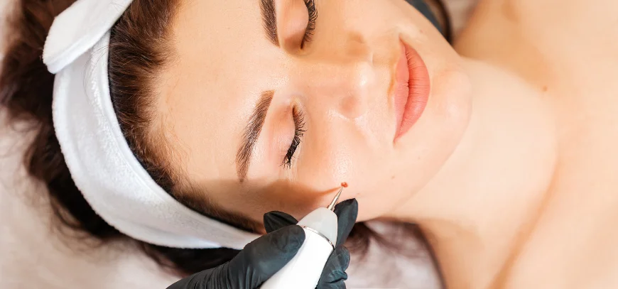
Overview
A mole, or melanocytic nevus, is a cluster of pigment cells. Many stay harmless and stable for years. Mole removal at AB Skin Clinic identifies the lesion first. Dr. S. Madhuri, trained in dermatology and dermatosurgery, then discusses a method. Not every dark spot is a mole, and not every mole should be treated with laser. Some lesions are better excised and, when the picture calls for it, examined under the microscope. The clinic does not biopsy every mole it sees.
Concerns this visit may address
This visit suits a raised mole on the neck or beard area that nicks during shaving, a smooth facial mole you would like reduced, or a mole under a strap that stays inflamed. Cosmetic concern is a valid reason to ask, once a clinician agrees the lesion is appropriate to remove. Change is a different concern. Growth, darkening, an irregular border, bleeding without a clear scrape, or persistent symptoms are reasons to seek prompt medical review. Only a clinician who can examine and, when needed, sample the lesion can make that judgement.
Who may benefit from an evaluation
Adults and adolescents with a bothersome but stable mole benefit from an exam that names the lesion before technique is discussed. People with many moles may want one or two addressed and the rest observed. Describe a change when you contact the clinic. Mention pregnancy, blood thinners, thick scars, and moles on the eyelid or lip, because those details change timing and method.
Possible contributing factors
Most moles appear in childhood and early adulthood under the influence of genetics and sun. A new benign mole can still appear later, so a new mark deserves a look. Pregnancy can darken moles slightly, and that change should still be reported. Prior incomplete removal, a scar through a mole, and home acids or threads can distort the border.
How a dermatologist evaluates the concern
The dermatologist asks when you noticed the mole, whether it has changed, and what it feels like. Lighting, dermoscopy, and comparison with your other moles help separate a stable nevus from a lesion that needs follow-up or a sample. If the story or the dermoscopic picture is concerning, the plan may be excision with histopathology rather than a cosmetic shave. The mole removal guide is useful reading; the in-person exam settles the diagnosis.
Treatment approaches that may be discussed
When removal is suitable, a dermatologist may discuss shave removal, excision with stitches, or laser only in selected cases. Shave removal can flatten a raised benign mole. Pigment can still recur at the base. Excision removes a full-thickness piece and is preferred when a complete specimen is needed or when a flat mole is changing. Laser is not a default. Vaporising a suspicious mole can remove the chance to study it. Histopathology is advised for some lesions and not for others, based on history, dermoscopy, and method. Wart removal follows a separate, viral pathway.
How the treatment works
Shave removal uses a blade to bring a numbed lesion level with the surrounding skin. The base may be smoothed with a method the dermatologist chooses. Excision takes the mole with a margin of normal skin when that is indicated, then stitches the wound. If a specimen is sent, microscopy can confirm or revise the clinical impression. Laser, when appropriate, is reserved for lesions where losing a specimen is acceptable. You should hear the likely scar before you consent.
What happens during a consultation or procedure
The first appointment is often examination and counselling. If the lesion is straightforward and you agree, removal may happen the same day or be scheduled. The skin is cleaned, the area is marked if needed, and local anaesthetic is injected. Stitches are removed later or left to dissolve, depending on the site. A dressing is applied and wound care is shown. Ask whether a specimen will be sent. Skin treatments cover many concerns; mole care stays on this medical path.
Preparation guidance
Arrive with clean skin and a list of medicines, especially aspirin, blood thinners, isotretinoin, and supplements that affect bleeding. If the mole is on the face, plan for a visible dressing. Avoid cutting, threading, or burning the lesion before review, because inflammation blurs the border. Hours are Monday to Saturday, 10:00 am–1:00 pm and 6:00 pm–8:30 pm; Sunday is closed. A mole that is changing quickly or bleeding without injury needs prompt clinical review.
Aftercare and recovery
Keep the dressing as advised, then cleanse gently and use the suggested ointment. Do not pick scabs. Non-dissolving stitches must come out on time; leaving them can mark the skin. Sun protection on a new scar for several months reduces darkening, which is common on the face and in darker skin tones. Report increasing pain, pus, fever, or a wound that opens. Colour can return after a shave if pigment cells remain at the base; that recurrence should be rechecked in clinic. If histology is pending, attend follow-up so the result is explained.
Risks, limitations, and realistic expectations
Every removal leaves a mark. The scar can be pink, pale, brown, raised, or stretched, especially on the chest, shoulders, and back. Infection, bleeding, stitch marks, and return of pigment are possible. Shave removal can leave mole cells below the surface. Excision trades that risk for a longer line and, when indicated, a specimen. Laser carries pigment and texture risk and is the wrong tool when a microscope diagnosis is required. The clinic will not promise scarless skin. A sound expectation is a correctly identified lesion and a scar you can care for as it matures.
What influences suitability and results
The clinical diagnosis is the main factor. A stable, bothersome raised mole is a different decision from an irregular or changing lesion. The back and chest often spread scars more than the face. Keloid history, current medicines, smoking, and size affect healing and closure. Skin tone influences how visible a pale or dark scar will be. Incomplete past removal makes the next procedure less predictable. A preference for laser cannot override the need for a specimen.
Why professional dermatology guidance matters
Moles sit between cosmetic preference and the need to detect lesions that should not be destroyed blindly. A thread, a home acid, or a salon tool cannot make that distinction. Removing a lesion without understanding it can leave a scar and can discard the chance to diagnose it. Dermatologists use dermoscopy and, when needed, histopathology so treatment follows the finding. If size, colour, border, or symptoms change, the useful response is a prompt examination.
Why consider AB Skin Clinic
AB Skin Clinic is led by Dr. S. Madhuri, MBBS, MD DVL, DNB DVL, Fellowship in Dermatosurgery and Lasers, with around ten years in dermatology. She is listed as a consultant at Apollo Hospital, Secunderabad, a former assistant professor at ASRAM, and a former senior resident at Siddhartha Medical College. Laser is used only when it fits the lesion. Consultations run at Triveni Towers, MIG-154, 1st floor, Road No. 1, KPHB Colony, Hyderabad 500072, 94949 46701, and at Second Floor, Sy No 140, BLOCK-A, Plot No 14, above HDFC Bank ATM, Alkapuri, near Neknampur, Gandipet, Telangana 500089, 98489 99917. Hours are Monday to Saturday, 10:00 am–1:00 pm and 6:00 pm–8:30 pm; Sunday is closed. The clinic states that USFDA-approved lasers are used. Book an appointment and say you want a mole assessed.